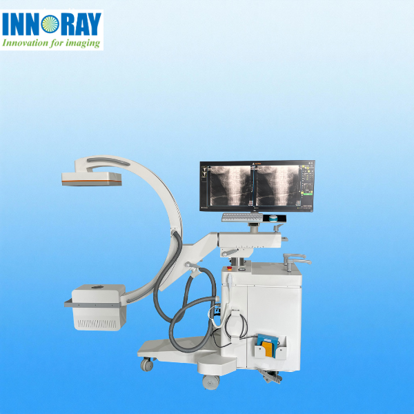
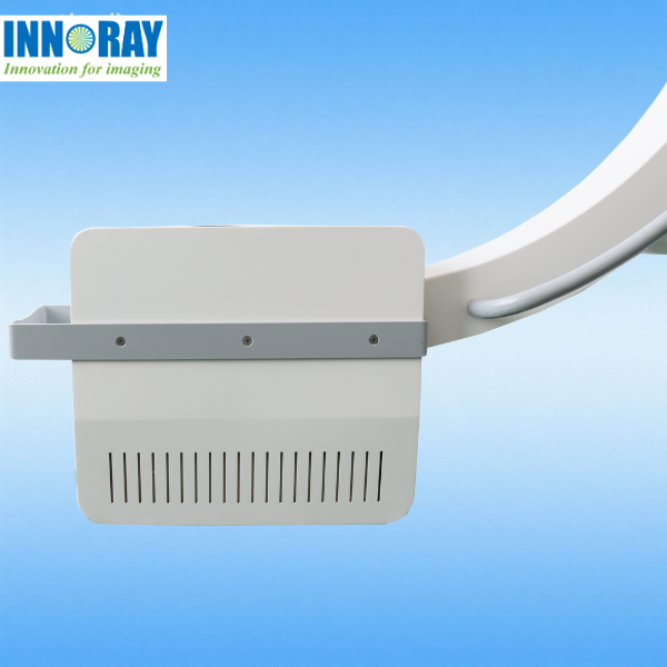
Adopts high-resolution flat-panel detector (FPD) with 16-bit grayscale output, delivering 2K+ image resolution (up to 2048×2048 pixels) for clear visualization of bone structures, soft tissues, and tiny lesions.
Supports real-time fluoroscopy (15-30 frames/second) and digital radiography (DR) mode switching, meeting needs for both dynamic observation (e.g., orthopedic reduction, catheter placement) and statici imaging.
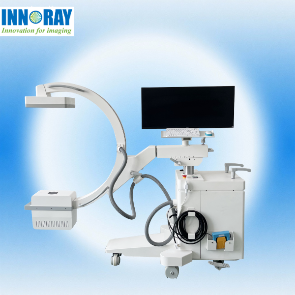
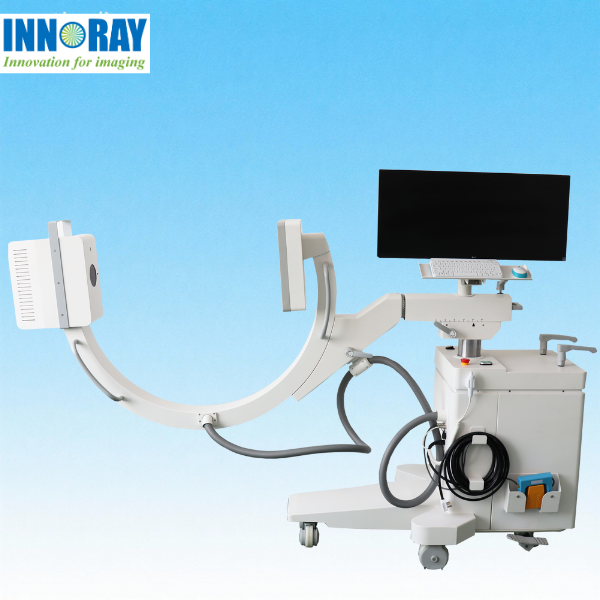
Compact and portable structure (wheelchair-mounted or floor-standing) with anti-collision sensors, suitable for operating rooms, emergency departments, clinics, and mobile medical scenarios.
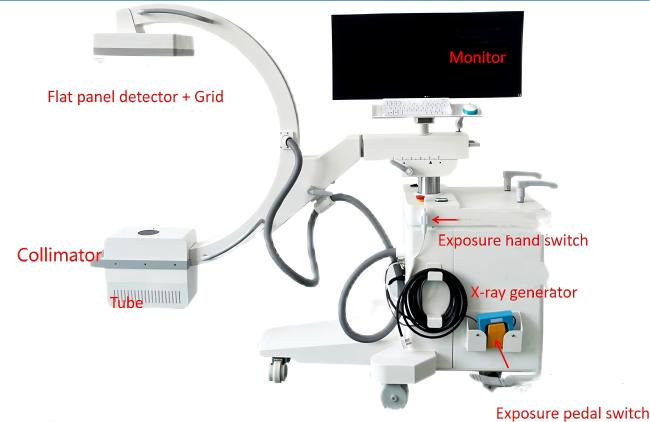
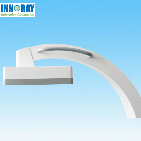
C-shaped arm with 180° rotational movement (horizontal/vertical) and adjustable focal length (50-120cm), enabling multi-angle imaging of different body parts (spine, limbs, chest, abdomen).
Compact and portable structure (wheelchair-mounted or floor-standing) with anti-collision sensors, suitable for operating rooms, emergency departments, clinics, and mobile medical scenarios.
Digital flat-panel detector replaces traditional image intensifiers, eliminating image distortion and improving spatial resolution and contrast ratio—critical for accurate clinical decision-making.
Real-time fluoroscopy with high frame rate ensures clear visualization of dynamic procedures, reducing surgical errors and repeat operations.
One-key start-up and rapid imaging (≤3 seconds per exposure), shortening procedure time and increasing patient throughput.
Intuitive touchscreen interface with customizable parameter presets (for different body parts/procedures) simplifies operation, reducing training costs for medical staff.
No need for film, developer, or fixer, reducing material costs and environmental pollution associated with traditional X-ray equipment.
Long service life of FPD (≥50,000 exposures) and low maintenance requirements, lowering total ownership costs (TCO).
Compact design fits in limited spaces (e.g., small operating rooms, clinics) and supports mobile use, expanding application scenarios beyond fixed radiology departments.
Compatible with various accessories: surgical tables, patient positioning aids, and specialized collimators (for pediatric/adult patients), enhancing clinical versatility.
Meets CE, FDA, ISO 13485, and other global medical device certifications, ensuring market access in Europe, North America, Asia, and other regions.
DICOM compatibility enables interoperability with existing hospital information systems, facilitating workflow integration and telemedicine collaboration.
Rigorous quality control during production, with shockproof, dustproof, and waterproof design (IP54 rating) for stable performance in harsh clinical environments.
Low radiation dose design aligns with global trends in radiation protection, enhancing patient safety and improving medical staff job satisfaction.
Ergonomic C-arm handle and adjustable height (70-150cm) reduce operator fatigue during long procedures.
Lightweight and easy-to-move structure (floor-standing models: ≤200kg) enables quick relocation between departments.


Application Scenarios of C-arm Series DR
1. Orthopedics
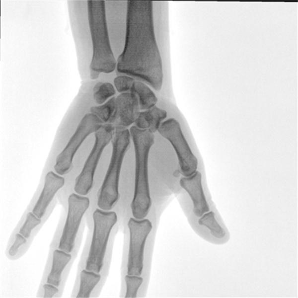
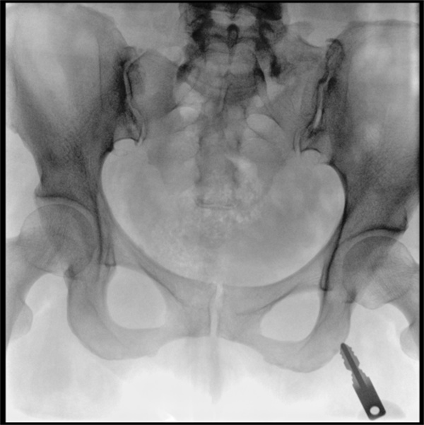
Fracture Treatment: Real-time fluoroscopy for closed reduction of limb, spinal, and pelvic fractures; guidance for internal fixation (screws, plates, nails) and external fixation device placement.
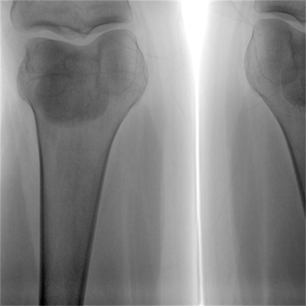
Joint Surgery: Intraoperative imaging during hip/knee replacement (component alignment verification), arthroscopic procedures (e.g., meniscus repair), and joint dislocation reduction.
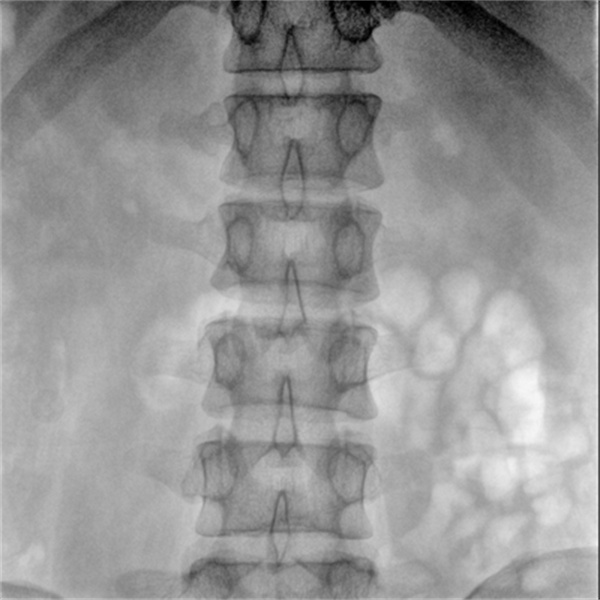
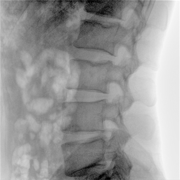
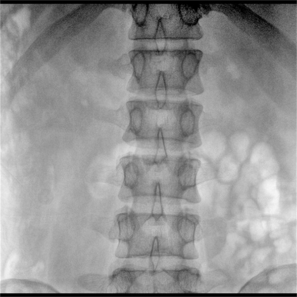
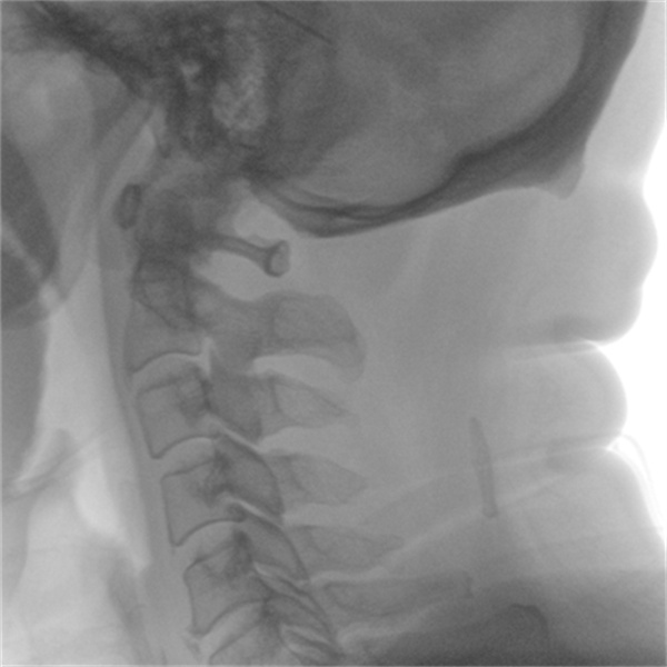
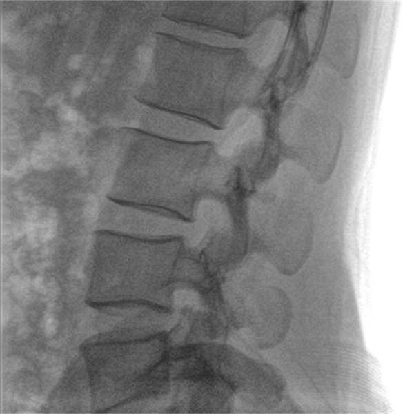
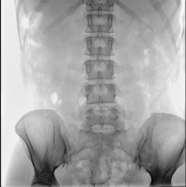
Spinal Surgery: Pedicle screw insertion guidance, intervertebral disc herniation surgery, spinal fusion monitoring, and scoliosis correction.
Pediatric Orthopedics: Low-dose imaging for pediatric fracture diagnosis and treatment (e.g., growth plate injury assessment) with adjustable collimation to fit small body sizes.
2. Interventional Radiology & Cardiology
Vascular Interventions: Catheterization (arterial/venous access), angioplasty, stenting, embolization, and thrombolysis (for stroke, pulmonary embolism, or peripheral artery disease).
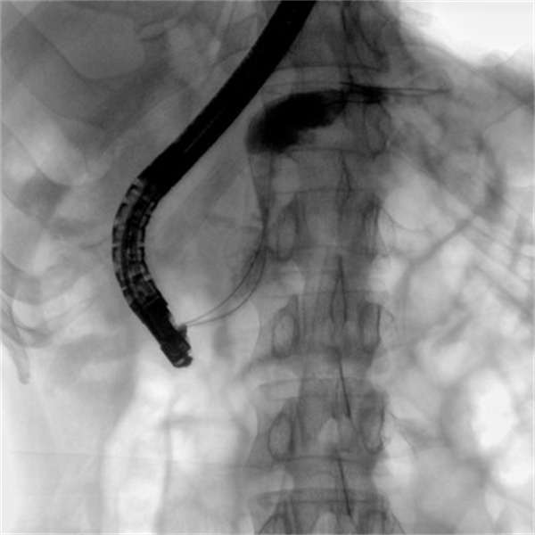
Non-Vascular Interventions: Biopsy guidance (liver, kidney, lung), abscess drainage, gastrostomy tube placement, and foreign body removal.
Cardiac Procedures: Pacemaker/defibrillator lead placement, coronary angiography, and balloon valvuloplasty.
3. Emergency Medicine
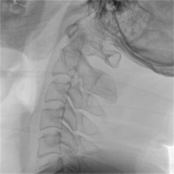
Trauma Diagnosis: Rapid bedside imaging for acute fractures (e.g., skull, rib, extremity), visceral injuries (liver/spleen laceration), and foreign body detection (e.g., bullets, metal fragments).
Critical Care Support: Bedside imaging for intubated patients (endotracheal tube position verification), chest infection diagnosis (pneumonia, pleural effusion), and abdominal emergency evaluation (bowel obstruction, perforation).
Point-of-Care Testing: Quick imaging in emergency departments, ambulances, or disaster sites to facilitate timely treatment decisions.
4. Operating Rooms (OR)
General Surgery: Intraoperative imaging for laparoscopic/open procedures (e.g., gallbladder surgery, hernia repair) to confirm anatomical structures or device placement.
Urology: Kidney stone removal (lithotripsy guidance), ureteral stent placement, and prostate surgery.
Gynecology/Obstetrics: Pelvic fracture management during childbirth, uterine fibroid embolization, and minimally invasive gynecological surgeries.
5. Primary Care & Clinics
Routine Radiography: Chest X-rays (respiratory infection screening), abdominal imaging (gastrointestinal disorders), and bone lesion detection (e.g., osteoporosis, tumors).
Sports Medicine: Diagnosis of sports-related injuries (sprains, strains, stress fractures) and rehabilitation progress monitoring.
Geriatric Care: Bedside imaging for elderly patients with limited mobility (e.g., hip fracture diagnosis, pneumonia screening) to avoid transfer risks.
6. Mobile Medical Services
Field Hospitals: Imaging support for disaster relief, military operations, or remote areas with limited medical resources.
Home Healthcare: Bedside imaging for homebound patients (e.g., chronic disease monitoring, post-surgical follow-up).
Mobile Clinics: Pop-up healthcare services in rural or underserved regions for basic radiological diagnosis.
7. Veterinary Medicine
Veterinary Orthopedics: Fracture reduction, joint surgery, and spinal procedures for small animals (dogs, cats) and large animals (horses, cattle).
Veterinary Emergency Care: Trauma assessment and foreign body detection in animals.